The oldest clinical case of the genetic condition that leaves men with smaller testicles has been discovered in the skeletal remains of a man who died 1,000 years ago in Portugal
- Skeletal remains showed that the man who died 1,000 years ago had Klinefelter syndrome, which is caused by an increase in the X chromosome.
- This is the oldest clinical case of an extremely rare genetic condition
- The skull features larger than average teeth, which is a width
- The remains were found in Portugal and buried in an oval tomb
The oldest clinical case of an extremely rare genetic disease that negatively affects testicular development has been revealed in the remains of a man who died 1,000 years ago in Portugal.
Known as Klinefelter syndrome, this condition gives men an extra X chromosome that greatly affects motor skills like standing and sitting, as well as their intellectual development – but it’s only present in about one in every 1,000 genetic male births.
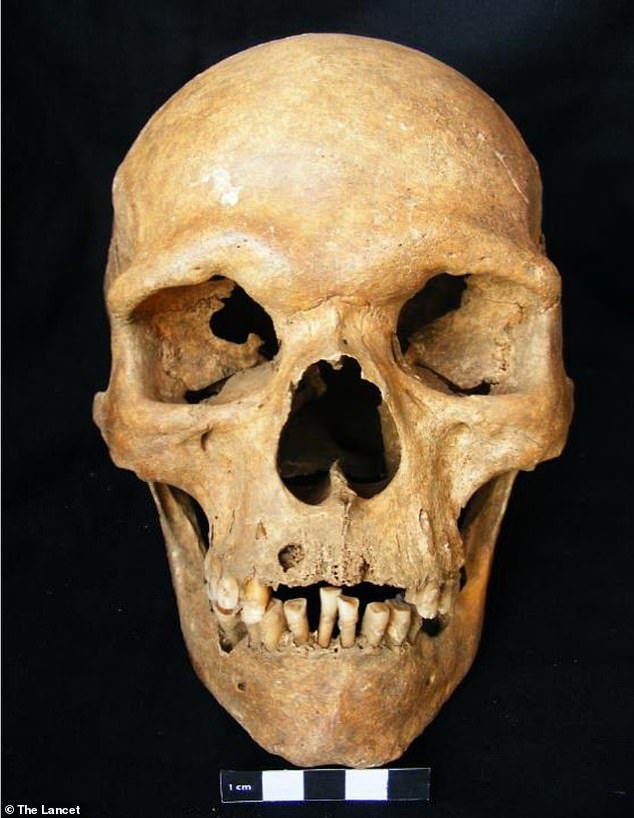
Although archaeologists didn’t know the remains were riddled with disease, the skull does show signs such as larger-than-average teeth and a serious gum infection that damaged the soft tissues in its mouth before death – both of which are caused by the genetic condition.
Klinefelter syndrome was first described in 1942, but the older condition, according to the researchers, provides a better understanding of how the disease developed through time and spread throughout history.
Scroll down for the video
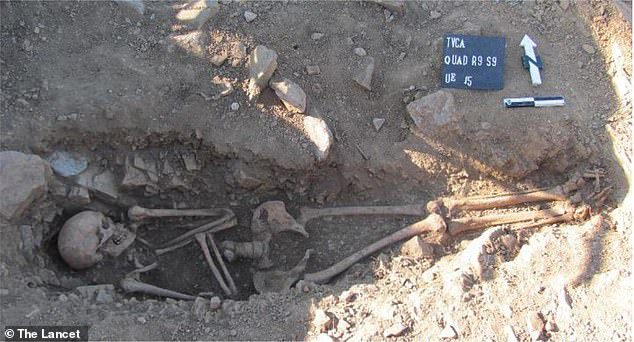
The 1,000-year-old skeleton was discovered in an elliptical tomb in Portugal. The team extracted DNA from the remains to reveal that the man had Klinefelter syndrome. Which happens when there is an extra X chromosome
The discovery, led by the Australian National University (ANU), was made while archaeologists were excavating the cemetery of Torre Velha, where they have found 59 graves so far.
The skeleton affected by the disease was buried in a single oval-shaped tomb, with no attached burial cap or goods.
He was placed in a supine position with the arms crossed over the chest, with the typical western and eastern orientation of a Christian burial.
The team began by analyzing genetic information obtained from a skeleton found in northeastern Portugal that was radiocarbon dating to the 11th century by researchers from the University of Coimbra in Portugal.
The team combined genetic, statistical, archaeological, and anthropological information to create a definitive diagnosis after collecting the remains from the grave
Dr Joao Teixeira of the Australian National University said in a statement: “We were immediately excited the first time we looked at the results.
However, ancient DNA is often degraded and of low quality and abundance, which means we were wary at first.
Associate Professor Bastian Lama, Head of Molecular Anthropology at the Australian Center for Ancient DNA, said: “In recent years, ancient DNA has helped rewrite the history of humans around the world. Our study demonstrates that it is now a valuable resource for biomedical research and the growing field of evolutionary medicine.
DNA was extracted by Ph.D. A student at the University of Adelaide, Xavier Roca-Rada, who said: “Genetic analysis was performed to computationally map degraded DNA fragments from the X and Y chromosomes to the reference human genome.”

During the burial, the man was placed in a lying position with arms held over his chest, with the typical western and eastern orientation of a Christian burial.
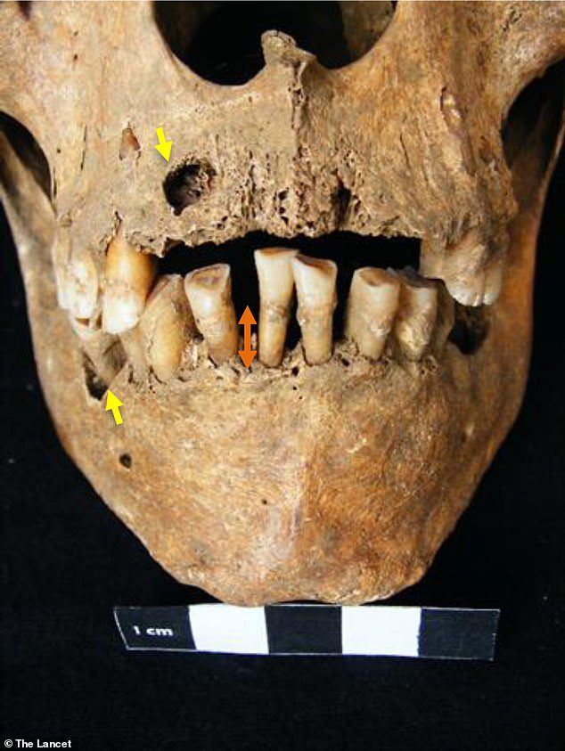
Although archaeologists did not know that the remains were riddled with disease, the skull features marks such as larger-than-average teeth and a serious gum infection that damaged the soft tissues in his mouth before death.
Given the well-preserved state of the specimen, the researchers were also able to identify physical features in the skeleton compatible with Klinefelter syndrome.
“Given the fragile state of DNA, we have developed a new statistical method that can take into account the characteristics of ancient DNA, and our observations to confirm the diagnosis,” said Dr. Teixeira.
While the study provides compelling evidence for the genetic history of Klinefelter syndrome, no social implications can be drawn from this diagnosis.
The researchers suggest that their new method of analyzing this particular skeleton could be further improved to study various chromosomal abnormalities in other archaeological samples, including Down syndrome.

Edith Wharton was a Pulitzer Prize-winning American novelist, short story writer, and designer best known for her insightful portrayals of upper-class society in the United States. Her works remain celebrated for their elegance, social commentary, and psychological depth.